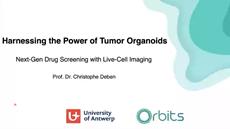
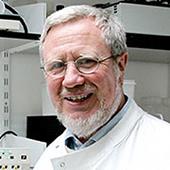
Dr. Christophe Deben
University of AntwerpAI-based analysis of 3D organoids – Accelerating translational cancer research
6 Apr 2023
3D organoid technology provides a unique opportunity to study health and disease biology in a biologically relevant setting, particularly for cancer research applications. Here, Dr. Christophe Deben, Group Leader of the University of Antwerp’s Tumoroid Screening Lab, shares his interest in organoid technology as a robust model for the translational study of cancer. He also explores how the University’s Center for Oncological Research (CORE) is using live-cell imaging of organoids to develop improved detection methods, screen new drug targets, predict therapy responses, and ultimately advance personalized medicine. Deben highlights how the use of AI-based image and data analysis software – specifically the CORE’s Organoid Brightfield Identification-based Therapy Screening (OrBITS) platform – enables researchers to automate their analysis processes, obtain deeper insights from live-cell images, and increase reliability, reproducibility, and throughput. This video was filmed at SLAS2023.
Christophe's Videos
Related Scientists


-

-
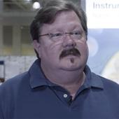
 Cancer Research Cell AnalysisDr. Kirk McManus Department of Biochemistry and Medical Genetics, University of Manitoba
 -
-

-

-
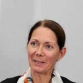
 Cell Culture Cell AnalysisDr. Theodossis Theodossiou Institute for Cancer Research, Oslo University Hospital
 -

-

-
-
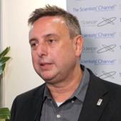
 Cell AnalysisProf. Stephen Hill Faculty of Medicine & Health Sciences, The University of Nottingham Medical School
 -
-sm.jpg)
-


-
-
-
 Cell AnalysisEli Bar, PhD Department of Neurological Surgery, Case Western Reserve University, Ohio
 -


Related Content